Saturday, September 3, 2022

PHYSIOTHERAPY TREATMENT FOR ADHESIVE CAPSULITIS


Adhesive capsulitis, also known as “frozen shoulder”, is a painful syndrome of the shoulder, which causes a progressive reduction in the range of motion of the shoulder, being one of the main causes of functional disability of the shoulder. 

One of the structures that make up the shoulder complex is the joint capsule. This collagen-based, elastic and flexible structure contributes to the stability and function of this joint.

Adhesive capsulitis is an inflammatory condition of the joint capsule that courses with contracture, pain and stiffness and promotes significant restriction of active and passive movement of the shoulder.

In many cases the cause is unknown, but it is known that it is more common in women (40 to 60 years old), it can also be related to diabetes mellitus, thyroid disorders, previous trauma and almost always with reports of a previous episode in the other shoulder.

Adhesive capsulitis is a pathology that has a beginning, middle and end cycle. This cycle is divided into 4 phases:

==>> Inflammatory phase: usually in the first 3 months. In this phase, the patient presents acute pain on active or passive movement and limitation for flexion, abduction and rotations.

==>> Freezing phase: occurs between the third and ninth month of the disease and the patient has chronic pain on movement, in addition to significant limitation of movement.

==>> Frozen shoulder: at this time, which usually occurs between the ninth and fifteenth month of the disease, the pain is minimal, but the limitation of movement (especially rotations) is quite significant.

==>> Thawing phase: last phase of the disease, the patient already has a considerable improvement in range of motion and the pain is almost zero.

The treatment of Adhesive Capsulitis is basically conservative, and the most important thing is to respect the patient’s symptoms in decision making. In the initial phase, the patient usually seeks a medical service and performs serial blocks of the subscapular nerve.

When arriving at physiotherapy, if the patient has a lot of pain and inflammatory signs, it is worth investing in resources such as combined therapy, laser, US and analgesic currents. The range of motion must be maintained and manual therapies with the aim of recovering them will be introduced after the blockade, as well as the gain in muscle and sensorimotor strength.

Physical Therapy in Adhesive Capsulitis

The goal of physical therapy in adhesive capsulitis is to eliminate discomfort and restore shoulder mobility and function. Considering the pathophysiology of adhesive capsulitis of the shoulder, there are several modalities of Physiotherapy in Dwarka. Each procedure is an integral part of the physiotherapy program and must be in accordance with the clinical aspects and the stage of the condition.

At first, cryotherapy is performed for 30 minutes two to three times a day, then transcutaneous electrical neurostimulation (TENS), with the aim of reducing pain and mobilization with pendular exercises and gentle passive mobilization of the shoulder, which can and should be repeated at home by the patient.

Although heat applications help to reduce pain, they are not necessarily the most indicated, due to some factors, passive or active mobilization is the most effective measure, as they significantly help to increase the range of motion.

Kinesiotherapy

Initially, inflammation control is sought in the treatment, for this it is necessary to eliminate any activity that may worsen the condition.

The use of analgesics and non-hormonal anti-inflammatory drugs can help with recovery, and intra-articular corticoid infiltration is a very indicated measure to reduce pain and consequently improve mobility.

Another treatment option is manipulation of the glenohumeral joint under anesthesia, aiming to reduce the period of joint stiffness, being a technique that favors the early restoration of movements and accelerates the return of function in the primary form.

This procedure, although indicated, should be done with caution as it can cause aggravation in patients with osteopenia in the humerus, osteoporosis and diabetes, in addition to other complications.

Analgesia makes it possible to perform Codmann pendular exercises for decoaptation, relaxation of muscle spasm, bringing pain relief and maintenance of minimum joint amplitude.

Stretching

Stretching is used to increase the length of the soft tissue structures that were affected by the problem and consequently shortened, which causes mobility deficit. This procedure makes it possible to increase the range of motion.

There are several stretching techniques that can be used, and they are determined by the specialist Physiotherapist in Dwarka based on the assessment of the patient’s condition and needs. We can cite as examples the posterior capsular stretching and stretching with the hands behind the back.

It is recommended that these exercises be practiced with the individual standing with the lumbar spine flexed at 90º. Movements are performed clockwise, counterclockwise, latero-lateral and anteroposterior.

It is important that, at the time of execution, the patient has the scapular muscles relaxed as much as possible, this allows the movements to extend as far as possible.

The professional physiotherapist in Dwarka can also guide the patient to reproduce the exercises at home with the help of a specific object and in an appropriate way. In general, a stick is used for this.

Stretching seeks to increase the length of shortened tissue structures thereby increasing range of motion. There are several stretching techniques that can be applied, which will be indicated by the professional according to the patient’s need.

Stretching exercises, whether active or passive, aim to restore movement in the affected limb, improving not only symptoms such as pain, but also your activities of daily living.

Cryotherapy

Cryotherapy is a treatment that uses cold for therapeutic treatment, very common for problems related to the musculoskeletal system, especially in the acute phases of inflammatory processes.

The cold applied to the affected limb helps to reduce the inflammatory process and helps to reduce edema, relieving pain. The explanation for this reaction is the effect of cold on nerve fibers, since it slows down impulse conduction, decreasing sensitivity.

Ultrasound

Ultrasound as a therapeutic method has been used due to its deep heating effects, which causes an increase in blood flow, and increases the speed of tissue repair and consequent improvement of the lesion. 

When ultrasound penetrates the body, it can have an effect on cells and tissues through two physical thermal and athermal mechanisms, in thermal effects the ultrasound travels through the tissues, where a part of it is absorbed, and this leads to generation of heat within the tissue, where they lead to pain relief, decreased joint stiffness and increased blood flow, being of fundamental importance in the acute phase.

In athermic effects, stimulation of tissue regeneration, soft tissue repair, increased blood flow in chronically ischemic tissues, protein synthesis and bone repair can be observed. It is assumed that one or more of the following physical mechanisms are involved in the production of these athermal effects, such as cavitation, acoustic currents and standing waves.

TENS

Tens or electrotherapy is a treatment that uses electrical current to improve injuries, promoting pain relief, as it normalizes circulation in the affected region, while activating the system that inhibits pain.

TENS (Transcutaneous Electrical Nerve Stimulation) is an analgesic current where peripheral nerves are stimulated through electrodes attached to the skin for therapeutic purposes.

The intensity of the current will depend on the parameters used during the treatment, according to what was directed by the physical therapist.

Regardless of the associated measure, Physiotherapy in Dwarka is an indispensable treatment in these cases, and should be started as soon as possible It is important to understand that treatment done properly and as soon as possible allows for a faster improvement and reduces the chances of a possible functional limitation of the member.

If you identify any of the symptoms listed above, look for a doctor immediately, he will certainly, in addition to requesting appropriate tests and prescribing pain relief medication, refer you to a Physiotherapist in Delhi for adequate treatment of the problem.

The procedures listed above must be performed by a specialized professional Physiotherapist in Delhi, and in the case of the use of anesthetics, follow-up by an anesthetic team.

Physiotherapy sessions in Dwarka for the treatment of Adhesive Capsulitis are usually prolonged and performed daily. For this reason, the understanding and cooperation of the patient in relation to the exercises is also a predominant and essential factor for the improvement and recovery of the patient.

Also remember to be careful when doing exercises and other activities that can cause injury to the muscles and joints of the body. Always seek the follow-up of a professional in the case of gyms for example.

Monday, August 29, 2022

Physiotherapy in temporomandibular joint dysfunctions


Jaw pain? Ringing in the ears? Dizziness? Neck and shoulder pain? Blockage when opening or closing the mouth?…

Many of these symptoms are caused by dysfunction of the temporomandibular joint.

Temporomandibular joint concept

When we talk about the temporomandibular joint, we refer to a joint that is made up of two bones, the temporal and the lower jaw.

They constitute, therefore, two joints that, like a hinge (one on each side of the face), connect the jaw with the skull. It allows us to move our mouth up and down and to the sides, making possible such important functions for the human being as digestion and speech.

Therefore, starting from this premise, we can imagine how much repercussion the dysfunction of said temporomandibular joint can have for a patient.

What is a temporomandibular dysfunction?

When we talk about temporomandibular dysfunction (TMD), we are referring to a functional type of alteration that causes pain around the TMJ and surrounding muscles.

This dysfunction affects all the structures that are related to and give shape to this joint, as is the case of the bony parts, muscles (such as the masseter) and the entire vascular-nervous package (nerves, arteries).

TMD can affect the ability to speak, eat, chew, swallow, make facial expressions, and even breathe.

According to the most current scientific evidence, a very high percentage of the population suffers or has suffered from a dysfunction at this level. There is a prevalence in the population between twenty and forty years of age and a higher incidence in women than in men.

Causes of TMJ dysfunctions

There are many factors that cause and generate TMJ dysfunction, thus being considered a disease of multifactorial origin.

Among the most frequent, we find:

  • Biomechanical causes: dental malocclusion, lack or loss of a dental piece and some dental treatments.
  • Psychological causes: Increased stress, anxiety; which leads to somatization and increased muscle tension at this level.

If you are so stressed that you clench your jaw and grind your teeth, you may develop temporomandibular joint (TMJ) related pain.

  • Metabolic type alterations.
  • Infections and autoimmune diseases.
  • Various forms of arthritis.

Symptoms of TMJ dysfunction

TMJ dysfunctions, due to the complexity of the joint itself, will generate a large number of symptoms, very different from each other. This can cause confusion and make an accurate diagnosis difficult.

For this reason, it is important to go to a qualified professional (dentist in Delhiphysiotherapist in Delhi expert in TMJ) to establish a good clinical diagnosis and propose the best treatment for each case.

As the most important symptoms of said injury, we find:

  • Local pain in the joint area.
  • Stiffness of the jaw muscles.
  • Pain radiating to the neck and shoulder area.
  • Clicking, crackling, or grinding sound of the TMJ when you open or close your mouth.
  • Headache or headaches, of great intensity, with the possibility of irradiation to the eye area.
  • Limitation in the functionality of the TMJ, with difficulty chewing, opening and/or closing.
  • Alterations at the hearing level, with the possibility of tinnitus or ringing in the ear, pain, dizziness and vertigo and even decreased hearing capacity.
  • Dysphagia or difficulty swallowing (trouble swallowing, making it difficult to eat).
  • Paresthesias or tingling of the entire facial area.
  • Tooth sensitivity.
  • Psychological alteration, depression and/or anxiety type.

Treatment of TMJ dysfunctions

As we have been able to observe, temporomandibular dysfunction can present with very different causes and symptoms in different regions of the body.

For this reason and given the complexity of the joint, it is necessary for the patient to receive joint treatment by a multidisciplinary team of specialists: dentist in Delhiphysiotherapist in Dwarka, neurologists, among others. In order to receive a comprehensive and integrated program of care.

Within physiotherapy in Dwarka, treatment begins with:

  1. A good clinical diagnosis.

To do this, a clinical history of the patient will be made, with a correct anamnesis. We will also perform neurodynamic tests where we will ask if you have pain in the joint, difficulty opening or closing your mouth, if you have joint sounds or clicks, headache or dizziness…

  1. Later we will carry out a complete evaluation of the joint: joint mobility and functional examination.

With all these data we establish:

  1. Most suitable treatment.

This will consist of:

  • Massage therapy techniques for all the muscles involved.
  • Joint mobilizations.
  • Stretches.
  • manipulative techniques.
  • Dry needling of muscle trigger points.
  • applied electrotherapy.
  • Home exercise guidelines.

All of this will help relax the affected muscles and tendons with consequent pain relief and improvement in range of motion.

It has been proven that after adequate physiotherapy treatment, the muscles of mastication and the neck achieve a more normal muscle tone, reducing signs and symptoms.

Conclusions

The temporomandibular joint (TMJ) is one of the most complex and most used anatomical structures in our body. It is responsible for chewing, swallowing and phonation and must work in a balanced and symmetrical way to achieve proper function.

In TMJ injuries, even more so, a good clinical diagnosis is extremely necessary. This will allow us to discern and rule out other more serious injuries that may be causing the pain in this region. Thus, later, we can establish a good treatment to improve function.

Interdisciplinary work is of the utmost importance. Only a coordinated and qualified team of physiotherapist in Dwarka will manage to treat your pathology with assured success.

Monday, August 22, 2022

MENISCUS TEAR AND PHYSICAL THERAPY


The most popular option is arthroscopy in Delhi for knee surgery, however, there are conservative treatments with physiotherapy in Dwarka, which speed up recovery times and strengthen the patient’s joint.

WHAT IS A MENISCUS TEAR?

The meniscus tear is one of the most common knee injuries along with the anterior cruciate ligament, especially among athletes. It is caused by a break or tear in the fibrocartilage, which acts as a cushion between the tibia and the femur, known as the meniscus.

The meniscus plays a fundamental role in the knee joint, as it helps distribute weight in the joint and directly affects its stability.

Causes of meniscus tear

Depending on the factor that causes the meniscus tear, there are different causes, however, they all have a common factor and that is that the tear generally occurs after a sudden movement of the knee.

In the first place, degenerative causes can be differentiated, in this case, the meniscus tear is caused by cartilage wear and tear, which is usually common in patients with degenerative diseases, such as osteoarthritis or osteoporosis. These breaks are common in older people.

On the other hand, traumatic breaks are differentiated, which are produced by an impact. And, finally, mechanical breakage, which is due to poor posture or joint disposition, which causes it to malfunction. These types of breaks are more common among athletes.

Symptoms of meniscus tear

The symptoms of a meniscal tear are very characteristic. First of all, it is common for the patient to have pain in the area which, in many cases, is accompanied by inflammation.

In addition, patients usually present immobility and joint weakness, which prevents them, in many cases, from performing usual activities, including, in some cases, walking.

Meniscus tear treatments

Once a meniscus tear has been diagnosed, the most common is for the patient to be prescribed anti-inflammatory treatment, with the aim of reducing inflammation and relieving pain.

In some cases, it is necessary to have the patient undergo a meniscus operation, in which the entire meniscus may be removed, or in other cases, the torn meniscus is sutured.

PHYSIOTHERAPY TREATMENT FOR MENISCUS TEAR

One of the most effective treatments for meniscus tears and fractures is physiotherapy in Delhi and knee rehabilitation, which can be applied at different times. In some cases, the rupture is treated primarily with physical therapy, to prevent the patient from undergoing surgery. In cases in which the patient is operated on, a post-surgery physiotherapy treatment in Dwarka is carried out to rehabilitate the joint. In order for the recovery time to be as short as possible, the operation must be performed by an orthopaedic surgeon in Delhi specialized in minimally invasive surgical techniques.

Physiotherapy treatment in Dwarka has numerous benefits, since it strengthens the joint, preventing its chronification, in addition, it contributes to the recovery of mobility and pain relief

Physiotherapy techniques for torn meniscus

Physiotherapist in Dwarka focuses on exercises to regain the strength and stability of the joint, with the aim that the patient can return to their usual activities. In addition, treatment with physiotherapy tries to avoid a sedentary lifestyle, so that the patient does not lose muscle capacity.

Some of the techniques used by the best physiotherapist in Dwarka is the release of adhesions from the joint tissues and draining massages for the evacuation and reabsorption of the inflammatory liquid.

In addition, in some cases electrostimulation is applied to reduce pain and inflammation. Through patches placed on the patient’s joint, electrical impulses are generated, with the aim of restoring the mobility of muscles, ligaments and tendons, and for the patient to recover muscle tone in a short period of time.

Physiotherapy treatments are recommended by physiotherapist in Delhi, because they work to strengthen the joint and restore mobility. For this, it is important that the specialist studies the patient’s case and designs a table of exercises adapted to the patient’s characteristics.

Physiotherapy exercises for torn meniscus

As mentioned, physiotherapist in Dwarka prevents the patient from losing muscle tone. It focuses on increasing muscle strength and proprioceptive exercises to work on balance, that is, those exercises that stimulate the nerve receptors found in the muscles and joints.

In addition, in many cases, gait re-education is carried out, with the aim of correcting those postures that involve forced positions for the joints and that can cause the patient to relapse into the injury.

BENEFITS OF TREATING MENISCAL TEARS WITH PHYSICAL THERAPY

As explained, physiotherapy treatments in cases of meniscus tears have very good results in patients. In addition, they are as effective as surgery and recovery times are shorter.

In addition, physiotherapist in Delhi focus on strengthening the joint, which means that the patient does not lose muscle tone and can almost fully recover their usual activities.

Wednesday, August 17, 2022

Physiotherapy treatment for patients with spinal cord injury


Spinal cord injury is a public health problem, which produces disability and dependency, physiotherapy plays a very important role in the treatment of patients with this type of condition.

What is spinal cord injury?

Spinal cord injury is defined as any pathological process of diverse etiology (accidents, illness, among others) that directly affects the spinal cord and that causes, therefore, an alteration of sensory, motor or autonomic functions below the level of the spinal cord injury.
 
In general, the patient with this type of injury is classified with tetra and according to the name of the limbs that are affected, thus indicating the level of injury, also distinguishing between the concepts of paresis and plegia, depending on whether or not there is preserved motor capacity, therefore, the deficit experienced by the patient will be conditioned by the extent of his injury, extension and involvement of the substance.

What is the treatment through physiotherapy for this type of alteration? 

The consequences of accidents that produce a spinal cord injury and its complications turn the user with this condition into a subject who undergoes multidisciplinary rehabilitation and constant control and treatment supervised by professionals.

In our physiotherapy clinic in Dwarka we have professionals who treat patients with this type of illness, allowing them to develop their activities satisfactorily and that they obtain the best recovery.

Physiotherapy in relation to pain control and trophic disorders will be based on improving muscle tone, avoiding muscle atrophy as much as possible and reducing or controlling the pain that may arise from the injury. Pains that are originated due to myofascial trigger points.

They are treated through myofascial release techniques, as well as assisted passive kinesitherapy in the extremities in order to relax the limbs and maintain function. In addition, physiotherapist in Delhi can apply to denervated muscles or those that are healthy. 

Electrotherapy makes it possible to avoid muscle atrophy and increase the strength of the muscles that are preserved, in addition to this it is also applied to patients with spasticity, stretching and electrotherapy with ultrasound, massage, among other techniques… Dry needling is also used, technique applied by physiotherapist in Dwarka. 

Regarding the control of respiratory infections, inspiratory muscle training shows numerous benefits, infections must be prevented through physiotherapy using mobilization and daily activation of the respiratory muscles, in addition to this, a plan is also applied in the different sessions. Sloped in order to facilitate the movement of mucus from the apical bases, in the same way respiratory exercises combined with assisted or passive active movements of the upper limbs are also performed in patients with decreased respiratory muscle strength.

Respiratory muscle training with technical strength and resistance exercises and airway clearance such as autogenous drainage, manual and electrical techniques, as well as manual chest compression, volume-enhancing techniques, and practical exercises are the most commonly used for this. Diaphragmatic and chest breathing exercises and the use of inspiratory and expiratory volume incentives are used.

Regarding the treatment for the control of the alterations of the renal, digestive and urinary system, the treatment should allow the patient to stand up even walking with the purpose of achieving a movement of the intestinal tract through the free suspension system, other techniques used For patients with paraplegia, abdominal massages, Valsalva maneuver, flexion of the upper trunk, among other exercises focused on improving the patient’s quality of life, are used to increase pressure at the abdominal level.

At the level of the renal and urinary system, the best physiotherapist in Delhi must work the pelvic floor muscles, so that it has the ability to recover its strength, stimulation is encouraged either with manual, electrical or electropure stimulation, in this way the patient with exercises of the pelvic floor can perform these movements autonomously.

People who suffer from this type of spinal cord injury can have greater functionality through physical therapy rehabilitation. In our clinic we have best physiotherapist in Dwarka who will address this and other types of pathologies, in addition to that, we work based on integrative physiotherapy in Dwarka, which is a methodology which is based on working the body as a whole, from a holistic view, where a large part of the injuries or pathologies that affect the musculoskeletal system are originated through an emotional factor or an incorrect diet that ends up affecting an organ in one way or another, causing not a disease but a dysfunction of the same, which has repercussions on a muscular, nervous or bony structure that affects the patient.

Friday, August 5, 2022

Muscle contracture: Definition, symptoms, treatment and prevention

Muscle contractures can affect the performance of the person who suffers from it, in this article we will talk about them and their treatment and prevention through physiotherapy in Dwarka.

What is muscle contracture?

Muscle contracture, as its name indicates, is a continuous and involuntary contraction of the muscle or some of its fibers that appear when making an effort, this manifests as a bulging of the area that develops pain and alterations in the normal function of the muscle.

Muscle contracture usually appears when the muscle performs an activity that is inappropriate in intensity or function, as well as when more weight is taken in the gym than it should be, contractures may appear or when a sustained effort is made over time without taking on so much weight, such as case of poor placement on the spinning bike.

Muscular contractures can appear at the moment in which an exercise is being performed or after…

It occurs because there is an accumulation of metabolism that causes inflammation or because there is not enough blood supply in the area, it can also be due to excessive fatigue of the fibers that at the end of the exercise see their relaxation capacity diminished.

The first method of intervention of a contracture is its prevention and for this a good warm-up must be carried out in order to prepare the muscle before the effort, a progressive programming in intensity of the loads also helps from less to more.

Muscle contracture as the main cause of back pain

In cases where back pain is triggered, the contracture appears essentially when the muscle is required to work more than it can perform and makes it intense and punctual, such as a sustained and less intense effort such as maintaining for hours an inadequate posture, on the other hand, some alterations of the spinal column or imbalance can favor certain muscle groups that are constantly working more than necessary, which predisposes them to contract the same thing that happens when the muscles lack power and it is required exert forces that exceed their capacity. In some scientific studies it has been shown that the paravertebral musculature is symmetrical on the left and right sides.

In patients who have undergone surgical interventions in the back area or who have suffered chronic back pain, the paravertebral muscles can atrophy up to 80% with respect to that of the healthy side, facilitating an asymmetric distribution of loads, muscular overload or distal and the appearance of new painful episodes. In this situation, it is essential to exercise appropriately for each specific case in order to counteract this tendency and avoid recurrence of crises.

However, muscle contracture can be triggered as a result of back pain regardless of the reason, back pain itself can cause muscle contracture due to a reflex mechanism, in these cases the contracture is not the main cause of pain, however, is an added factor which can aggravate. The contracture can worsen some of its causes, for example, in a painful herniated disc, the muscular contracture can increase the compression force on the disc and thus facilitate the output of the nucleus pulposus.


What is the symptomatology of muscle contracture?

The characteristic and common symptoms of muscle contracture are pain and limitation of movement, both parameters offer a wide range of severity because in some cases the contractures go from being minor annoyances without impediment to completely disabling injuries.

The symptomatology revolves around two parameters: the affected area and the extension of the contracture. Focusing on the first parameter, we find, for example, that in upper regions of the body, such as the cervical area, the contractures can trigger symptoms truly aggressive such as dizziness, vertigo and migraine, a scalene syndrome, that is, the entrapment of the neurovascular bundle of the neck due to inflammation or contracture of the muscle belly of the scalene causes a feeling of heaviness in the arm, deep pain, paresthesia and hands cold.

A pyramidal muscle that increases its thickness due to a contracture can compress the adjacent structures and cause false sciatica...

In relation to the second parameter, the extension of the contracture, it is evident that a greater dimension of the contracture can aggravate the effects mentioned above.

How is a muscle contracture diagnosed?

A correct diagnosis interprets the symptoms manifested by the patient, later the diagnosis focuses on the palpation of the affected area in search of muscular bulges with greater tension, a muscular rod offers a certain resistance to palpation while the fingers slide over it and finding a point with greater resistance means that that specific point of the muscle fibers is contracted. This assessment, together with the pain signal caused to the patient, palpation of this muscle or this muscle point will confirm the presence of a muscle contracture.

The injured muscle does not achieve the same great mobility as the healthy muscle on the opposite side…

What are the causes of muscle contracture?

The contracture may be caused by any of the following problems: brain and nervous system disorders with cerebral palsy or stroke, hereditary disorders such as muscular dystrophy, nerve damage, reduced use (such as lack of mobility) and scarring after a traumatic injury or burn.

From the point of view of oriental medicine, a theory applied in the Fiit concept, this type of alteration can also be triggered not only by physical factors, but also by emotional factors such as those related to stress (such as tension headache) or people. nervous and visceral factors that can trigger alterations in the stomach causing gastritis, this can also be caused by a poor diet such as excess coffee, excess fat, among others.

What is the treatment of muscle contracture?

In the first place, it is necessary to move away from the injury mechanism, if an exercise or the execution of a gesture triggers a muscular contracture, it is necessary to avoid executing them, relative rest is often the best method of healing and when we refer to relative is that the patient does not remain in very long periods of immobilization, but also remains active, because immobilization can cause adverse effects.

A treatment for the first stages of the contracture is the application of heat on the affected area because this provides vasodilation and contributes to the blood purification of the muscle segment, adding this causes a sensation of relief thanks to its analgesic and relaxing effect.

The massage therapy applied in our physiotherapy clinic in Dwarka is an effective and natural method because it allows us to eliminate the ailments that this alteration triggers.

From the point of view of integrative physiotherapy, assuming that the origin of this alteration may be due to emotional factors, it is advisable to reduce the levels of stress that may occur in people. These emotional factors can trigger contractures, therefore, we must work to reduce their presence as well as reduce the consumption of foods that cause irritability in the digestive segment, such as excessive consumption of coffee or high-fat foods.

Therefore, adequate nutrition is recommended by physiotherapist in Delhi in order to reduce or prevent the onset of muscle contracture...

How can a muscle contracture be prevented?

Whether it is because the patient has just come out of a contracture or because he has a tendency to suffer, the best way to prevent them and avoid relapses is by taking into account the following points:

  • Avoid repetitive gestures or movements, if the contracture is produced due to a sustained contraction over time, a constant repetition of a contraction favors its appearance, if it is unavoidable to repeat a gesture for sports or work reasons, it is convenient to carry out small routines of stretching and joint mobility exercises that last between 5 and 10 minutes in the areas exposed to the injury, this routine would be repeated periodically throughout the working day or in a training session, for example, a routine of 5 minutes every 2 work hours.
  • Adopt correct postural hygiene, whether while studying, watching television, in the office chair or even at bedtime. A bad posture can trigger contractions continuously, the longer the adoption of an incorrect posture the greater the contracture will be, in addition to adopting correct postures, a material must be used that guarantees its principles such as the use of ergonomic chairs at work time or a quality mattress for rest. 
  • Before undertaking any physical activity, it is essential to warm up according to the effort to be made. Acquiring the right muscle temperature through exercise is the best way to prevent possible injury.

This article explained a little about this alteration, its possible causes, treatment and prevention. If you present recurrent muscular contractures, visit our physiotherapy and osteopathy clinic we have experienced physiotherapist in Dwarka who will satisfactorily address these alterations to improve your health and quality of life.

Friday, July 29, 2022

PHYSIOTHERAPY AS A TECHNIQUE FOR THE RECOVERY OF OSTEOPOROSIS


Osteoporosis is usually associated with bone aging, but currently, poor lifestyle habits also lead to the appearance of this disease. It usually occurs in women around the time of menopause. In this article we tell you what osteoporosis is, its symptoms and causes, and how physiotherapy treatment in Dwarka helps in osteoporosis.

WHAT IS OSTEOPOROSIS?

Osteoporosis is a disease that weakens the bones, so that there is a decrease in bone mass density.

The word osteoporosis means “porous or leaky bone”.

Who is affected by osteoporosis?

Osteoporosis mainly affects women. There is a certain relationship between the lack of estrogen after menopause and the development of this disease. Thus, when hormone levels are low and there are few periods of menstruation, a loss of bone mass density is caused.

Therefore, women over 50 years of age are the risk group for osteoporosis.

Types of osteoporosis

There are several types of the disease that weakens the bones.

Primary osteoporosis is the most common type of osteoporosis. As already mentioned, it is due to the rapid loss of bone mass after menopause. This makes it a common disease in women, generally between the ages of 45 and 55 years old.

On the other hand, secondary osteoporosis presents the same symptomatology as the previous one. It has the particularity that it can appear at any age and can be caused by the intake of medications that cause bone decomposition.

Osteogenesis imperfecta is a particular form of osteoporosis that occurs at birth, so that bones break easily and, in many cases, for no reason.

Finally, idiopathic juvenile osteoporosis (IJO) appears between 8 and 14 years of age and is characterized by pain in the back and extremities, as well as the presence of fractures.

Symptoms of osteoporosis

Osteoporosis is also known as a silent epidemic, since in the early stages there are no symptoms.

As the bones weaken, the symptoms evolve, being characterized by:

  • Bone fragility.
  • Vertebral fractures.
  • Back pain and back deformities.
  • Back and stooped posture.
  • Loss of height due to crushing of the vertebrae.

Causes of osteoporosis

Sometimes, the appearance of this disease happens without a known cause. In general terms, age, and therefore aging and bone loss, cause the manifestation of osteoporosis.

As already mentioned, menopause accelerates bone loss, which multiplies the risk of suffering from osteoporosis.

The intake of alcohol and tobacco and a sedentary lifestyle are other of the most frequent causes of osteoporosis.

BENEFITS OF PHYSIOTHERAPY FOR THE TREATMENT OF OSTEOPOROSIS

The treatment of osteoporosis through physiotherapy in Dwarka plays an important role, in a way that favors the density of bone tissue.

It is essential to put yourself in the hands of Best Physiotherapist in Dwarka to see the results. One technique is magnetic therapy for the treatment of osteoporosis. The magnetic fields will help generate low and high intensity currents in the bone tissue, thus increasing the density of bone mass and helping bone recalcification.

As a key point to prevent osteoporosis, it is recommended to have adequate lifestyle habits, such as postural education, moderate physical exercise and a diet rich in calcium, suggests the top physiotherapist in Dwarka.

Exercises to strengthen bones

Exercising can help prevent osteoporosis. Muscle strengthening for osteoporosis is a clear example of this. Using weights and resistance bands, muscle groups can be strengthened, just like the muscles of the spine.

On the other hand, aerobic activities such as dancing or aerobic training machines benefit the bones, in addition to improving cardiovascular health.

Training flexibility with osteoporosis helps maintain good muscle function. Therefore, it is important to move the joints in their full range of motion.

In addition, performing exercises for balance and stability is a key point. This helps prevent falls while improving stability and balance.

Now that you know what the main causes of osteoporosis are and what its best treatment is, you will have been able to verify that the practice of sports exercise in patients with osteoporosis is a way of staying active in a safe way that also helps prevent the onset of osteoporosis. of osteoporosis. At Physiotherapy centre in Dwarka we have a team of specialists in physiotherapy and rehabilitation who can help you in the treatment of osteoporosis. Make an appointment at our Physiotherapy clinic in Dwarka and put yourself in the hands of our best physiotherapist in Delhi.

Thursday, July 21, 2022

WHAT IS ANKLE SPRAIN? FIND OUT HOW TO TREAT IT!

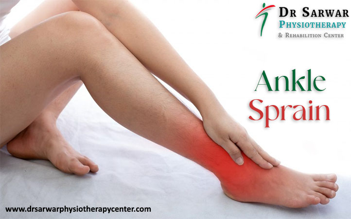
ankle-sprain

You know that sharp pain, that happens after an ankle sprain? An ankle sprain is a sudden movement, which leads to a strain or tear of ligaments in a joint. This type of sprain is the most common injury in the universe of musculoskeletal pathology.

The ankle is the joint between the foot and the leg and is made up of 3 bones: tibia, fibula, and talus. Patients tend to disregard these injuries, which can cause local symptoms and even permanent sequelae.

Ankle sprain is a common cause in young adults (ages 15 to 35) and athletes. According to physiotherapy doctor in Dwarka, ankle sprain is one of the most frequent diseases in orthopedic emergencies. It occurs most frequently in soccer, basketball, volleyball and athletes, accounting for about 10% to 15% of all sports injuries.

In an ankle sprain, the anatomical structures of the joint are usually affected, such as muscles, tendons and, in more severe cases, bones and cartilage, which can progress to ligament complications.

In a normal state, the ligaments, which are elastic structures, stretch to their limit, then return to their initial position. Ligaments have a specific range of motion that allow joint stability.

Ligaments are made up of whitish fibrous connective tissue (contains collagen) and have the function of joining two or more bones, protecting, and stabilizing the body’s joints and acting as shock absorbers.

In this text we will present everything you need to know about an ankle sprain, the main causes, its symptoms and the most appropriate form of treatment with promising results. Keep reading!

Classification of Ligament Injuries

Depending on how affected the ligament is and the degree of damage, an ankle sprain can range from mild to severe. Pain and swelling are common; however, it is possible to walk with little pain. In a more severe sprain, tenderness and bruising around the ankle may occur, causing difficulty in walking.

Grade I (mild): ligament strain occurs. Generally, the patient should feel improvement after three days, with asymptomatic or little tissue pain.

Grade II (moderate): partial tear with mild laxity and moderate pain, instability, swelling, and tenderness.

Grade III (severe): complete rupture, severe pain, with local swelling, significant laxity and, most of the time, the joint is unstable.

How an Ankle Injury Occurs

In ankle injuries, there is an accidental inversion of the foot with plantar flexion, especially when stepping on an irregular step or surface, using unstable shoes, such as high heels or even falling from one’s own height.

This anomalous movement causes an injury that starts in the anterior talofibular ligament, and can progress to an injury to the calcaneofibular ligament, with the increase in the energy of the trauma.

Signs and Symptoms of Ankle Sprain

In ankle sprain, pain occurs in the foot, from mild to very severe.

Usually, the ankle immediately begins to swell, and ecchymosis and joint effusion may develop in the area of ​​swelling. The pain increases with movement and is tender to the touch.

The more severe the injury, the more visible the signs.

What are the main causes of Ankle Sprain?

There are many causes, and some factors may involve the appearance of the lesion, such as:

  • sudden movements with the feet;
  • Wearing inappropriate footwear, such as very high heels;
  • Carrying out physical activity without preparation;
  • loose ligaments;
  • Walking on uneven ground;
  • Weak muscles;
  • Nerve injuries;
  • Previous injuries.

Diagnosis

The orthopedic professional usually performs the medical diagnosis of ankle sprain, as it is the most suitable to deliver the result and carry out a reliable analysis of the injury, through the patient’s clinical history, complementary exams that help in the exact and precise diagnosis of the injury.

Some of them are:

  • X-ray: allows the orthopaedic doctor in Delhi to obtain images with details of the region where the injury occurred, helping the specialist to exclude the possibility of a fracture in any bone of the foot or located in the ankle itself, thus allowing an assertive and complete diagnosis;
  • Magnetic Resonance Imaging: the specialist requests it when there is a more serious injury, which involves fractures in the articular bone;
  • Ultrasonography: allows observation of the ankle ligament while moving it, making it possible to visualize details of the joint injury;
  • Tomography: useful for a better assessment of the fracture pattern.

Types of Treatment for Ankle Sprains

Physiotherapy in Dwarka is the most effective method of treatment for sprains, especially when it comes to proprioceptive re-education, as it will prevent instability and new fractures.

Treatment can be either conservative or surgical. The choice depends on the severity of the injury. Conservative treatment will depend on the type and severity of the trauma.

The conservative protocol follows:

  • Rest: Do not support with the fractured limb;
  • Ice: Place on the injury site for 15 minutes every 2 hours for the first few days;
  • Compression: Bandages or elastic stockings help control edema;
  • Elevation: Keep the affected limb at heart level;
  • Immobilization: Immobilizing boot or plastered splint always in the initial care;
  • Medications: To control swelling and relieve pain in the first few days of trauma, such as anti-inflammatories and analgesics.

In the minority of cases, the treatment is surgical. However, depending on the type of fracture, more than one surgical intervention, called ligament repair, may be required.

When surgery is performed in the chronic phase, with patients with a history of repeated sprains, it is usually performed by fixing the injured ankle ligament to the bones, using implants such as anchors, which can be metallic or made of bioabsorbable material.

Conclusion

It is noted that from the articles studied there are different types of treatment for ankle injuries , such as the use of splints, immobilization with functional orthosis, elastic bandage, and others. The intervention criterion after an ankle sprain can be chosen according to the time of the event.

It is likely that there is an understanding in the literature when selecting the best practices for the treatment of injuries in the region, an understanding that assesses that the faster the choice of treatment type, the faster the patient’s recovery, says the best physiotherapist in Dwarka.

Monday, July 18, 2022

Muscle stretching: What to do when you suffer from the classic 'pull'

Muscle injuries are the most common type of injury in an athlete. In this article we want to focus on muscle elongation or muscle pull, the mildest muscle type injury, a priori.

Muscle strain is the famous “pull”. As Dr Sarwar, physiotherapist in Delhi, explains, it occurs when the muscle is stretched “beyond its possibilities”. It is usually considered the previous step to a muscle rupture and is considered a minor injury. "It is the most benign of muscle injuries," says Dr Sarwar, physiotherapy doctor in Delhi.

What is muscle stretching

Elongation occurs as a result of eccentric muscle contraction at the two ends of the muscle. It normally occurs in polyarticular muscles, such as the quadriceps and hamstrings. The fundamental difference with the break is that in the elongation the fiber does not break, or a minimal break occurs. “The rupture produces a hematoma, which takes longer to reabsorb and always leaves a scar, larger or smaller”, explains the physiotherapist in Dwarka. In contrast, stretching does not leave a scar, so the muscle does not lose functionality.

"It always occurs as a function of muscular overloads and imbalances ", illustrates the doctor. There are very characteristic examples. For example, in a short race: the explosive contraction of the quadriceps can produce an excessive lengthening of the hamstring. In soccer players it can occur when hitting the ball with force, which produces a sudden stretching of the posterior thigh muscles.

The sport most affected by muscle strain or pull is precisely football. "It generally occurs in the hamstring," explains the best physiotherapist in Dwarka, "followed by the rectus femoris, the adductors, and the twins."


When does a pulled muscle occur?

The elongation has several characteristics:

  • It occurs as a consequence of physical activity. It does not appear at rest but is the consequence of an eccentric contraction that is a little stronger than usual.
  • It usually appears when the muscles have not been properly warmed up.
  • Sometimes it occurs when explosive work is required of a previously excessively fatigued muscle. It is more frequent in the first phase of the season, when few training sessions have been carried out, or at the end, when fatigue accumulates.
  • External factors may influence. The case of excessive heat, humidity, a change in food or hydration defects do not help.

Symptoms of muscle strain are mild. The pain is usually diffuse, so that it is difficult to locate the exact point. Unlike a muscle tear, which usually generates rapid functional impotence, elongation can allow practicing sports, although with less intensity. "You can tell that something has happened, and sometimes it is not distinguishable from an overload because it is not excessively painful," Dr Sarwar abounds.

How to prevent muscle strains

Elongation is a type of injury that can be largely prevented, as long as some basic guidelines are taken into account:

  • Perform eccentric muscular work. "In any muscle injury, eccentric strengthening work is important, which improves flexibility and contraction capacity," says Albaladejo.
  • Pay attention to muscle rebalancing. Dr. Sarwar comments that it is common to find athletes with the quadriceps and little hamstring, and vice versa. "This ratio is very important to have balanced," physiotherapy doctor in Dwarka warns.
  • Do a good job of dynamic stretching in the warm-up phase. This will serve to increase your body temperature and your muscles' range of motion. Recent studies seem to indicate that stretching prior to exertion weakens the muscles and exposes them more to injury, as we explain in this article. For this reason, stretching should be accompanied by a movement that warms up the muscles.
  • Good hydration and a good diet. Low hydration hinders the transport of minerals, through which the brain sends electrical impulses that muscle cells transform into movement. Dehydration can cause muscle spasms, and these lead to the dreaded cramps or elongation of the fibers.
  • Avoid overtraining. Especially if you are returning to sports activity after a long period. Overtraining is reflected in muscle pain because the muscles need rest to recover and regenerate. Contractures, pulls or cramps can be related to excess. It can also cause muscle tears or tendinopathies.

What to do when you have a pulled muscle

Despite the apparent lightness of the muscle strain, it is advisable to stop practicing sports for a few days.

The treatment can be carried out in two phases:

  • The first two or three days it is convenient to control the inflammation by applying ice, in several daily sessions of twenty to thirty minutes. "Studies agree that in the first three days what you have to do is rest, ice, elevation and compression," says the physiotherapist in Delhi. In this way, the bleeding of the possible microtear is limited, which will reduce the scar.
  • As soon as the pain allows it, generally after the third day, you can move on to an activation phase. The goal is "to activate the circulation, begin to vascularize and work on strengthening with a smooth and controlled progression," says Dr Sarwar. Isometric contraction can also be worked on. That is, the contraction of the muscle without altering its length.

"Properly diagnosed, it should not take more than ten days to return to normal activity," concludes the physiotherapist in Dwarka.